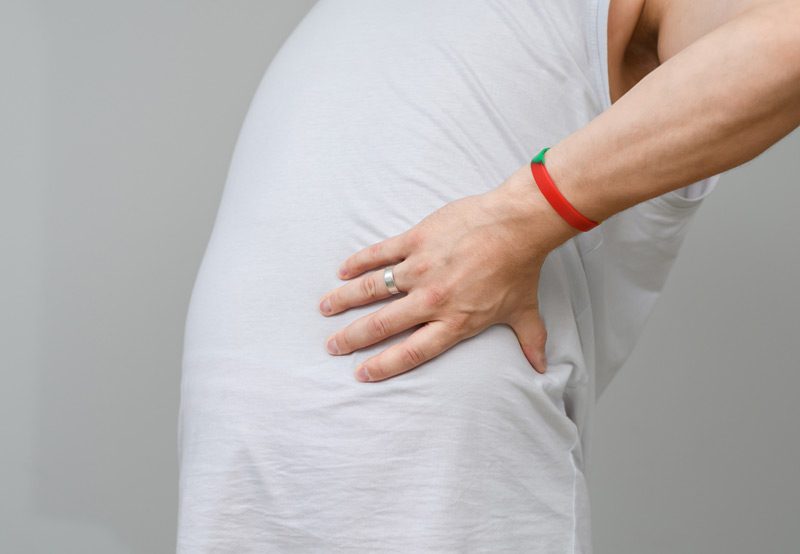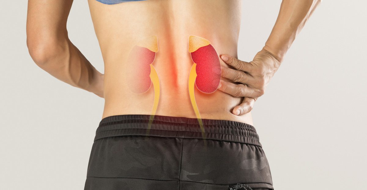

Renal colic is a type of severe, cramping pain caused by the sudden obstruction of urine flow—most often due to kidney stones blocking part of the urinary tract. The pain typically originates in the flank (side) or lower back and can radiate to the lower abdomen or groin. Renal colic is considered a medical emergency due to the intensity of discomfort and the potential for complications if left untreated.
Causes of Renal Colic
The most common cause of renal colic is a kidney stone becoming lodged in the ureter, the narrow tube that carries urine from the kidney to the bladder. As the body attempts to pass the stone, spasms in the ureter lead to sharp, intermittent pain. Contributing factors include:
- Dehydration: Low fluid intake concentrates urine, increasing the likelihood of stone formation.
- Dietary Factors: High sodium, animal protein, or oxalate-rich foods may promote stone development.
- Family or Personal History: A history of kidney stones increases the risk of recurrence.
- Metabolic Disorders: Conditions that affect calcium or uric acid levels can contribute to stone formation.
- Urinary Tract Abnormalities: Structural or functional issues in the urinary tract can increase the risk of blockage.

Symptoms
Renal colic is characterized by intense, often wave-like pain. Common symptoms include:
- Sudden, severe pain in the back, side, or lower abdomen
- Pain that may radiate to the groin or genital area
- Nausea and vomiting
- Hematuria (blood in the urine)
- Frequent or urgent need to urinate
- Painful urination
The pain may come in waves and fluctuate in intensity, corresponding to the movement of the stone within the urinary tract.
Diagnosis
Prompt diagnosis helps prevent complications and guide appropriate treatment. A urologist may use the following tools:
- Medical History and Physical Exam: To assess pain characteristics and rule out other causes.
- Urinalysis: To detect blood, infection, or crystals in the urine.
- Blood Tests: To evaluate kidney function and signs of infection or elevated calcium.
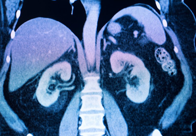
- Imaging: A non-contrast CT scan is the gold standard for detecting stones. Ultrasound or X-ray may also be used, depending on the situation.
Treatment Options
Management depends on the stone’s size, location, and the severity of symptoms:
- Pain Control: NSAIDs and narcotics are commonly used for immediate relief.
- Medical Expulsive Therapy: Alpha-blockers like tamsulosin can help relax the ureter and facilitate stone passage.
- Hydration: Increased fluid intake may aid in flushing out the stone.
- Surgical Intervention: If the stone is too large or causing complications, options include:
- Ureteroscopy with Laser Lithotripsy: A scope is used to break and remove the stone.
- Shockwave Lithotripsy (SWL): External sound waves fragment the stone.
- Percutaneous Nephrolithotomy (PCNL): For larger or more complex stones, this minimally invasive procedure accesses the kidney directly.
Next Steps
If you experience sudden, severe flank or abdominal pain, especially with nausea or blood in the urine, seek immediate medical attention. A urologist can determine the cause and guide you through treatment and prevention strategies to reduce the risk of recurrence.
