Dr. Mohammed Shahait
General Urology & Endourology Specialist
BOOK APPOINTMENT

Meet
Dr. Mohammed D. Shahait
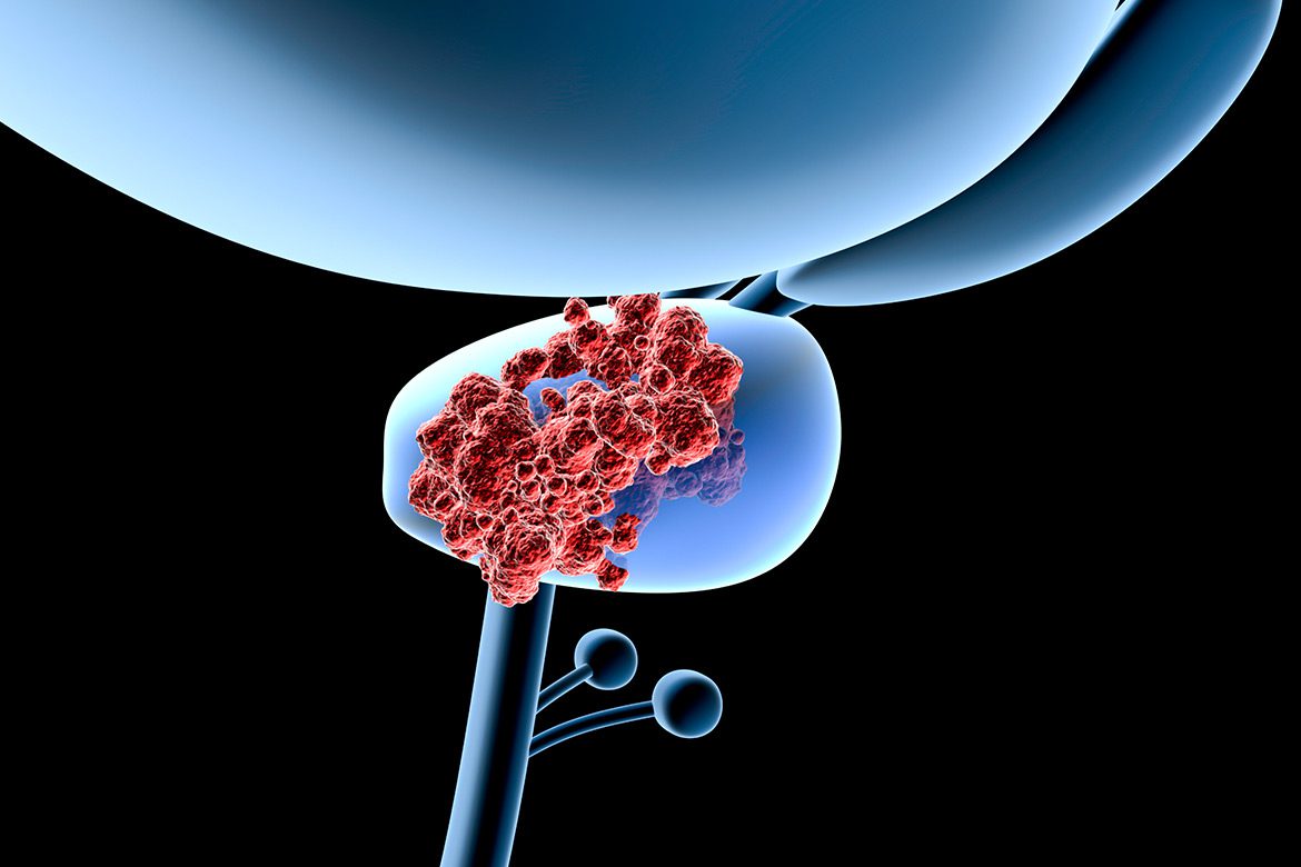
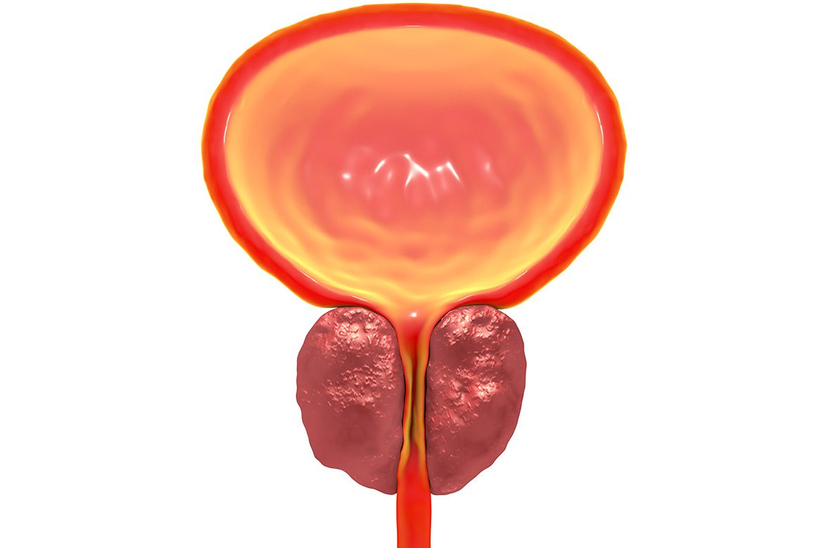
Dr. Mohammed Shahait is a dedicated urologist with a special interest in the diagnosis and treatment of prostate diseases, including benign prostatic hyperplasia (BPH), elevated PSA, and prostate cancer. As a prostate disease specialist, he utilizes advanced diagnostic techniques, such as MRI-guided transperineal prostate biopsy, ensuring precise and minimally invasive evaluation of prostate conditions.
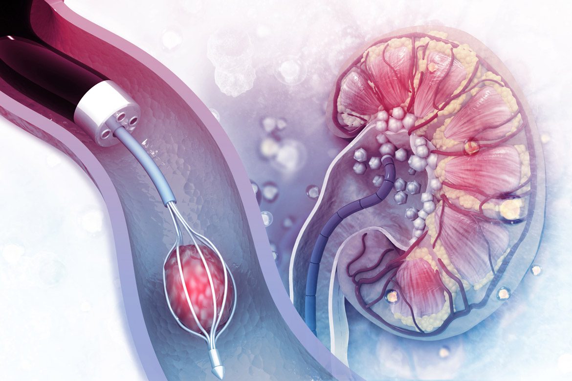
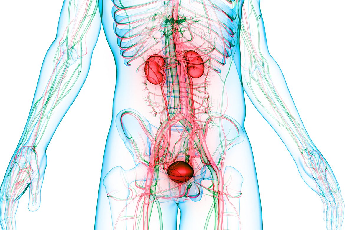
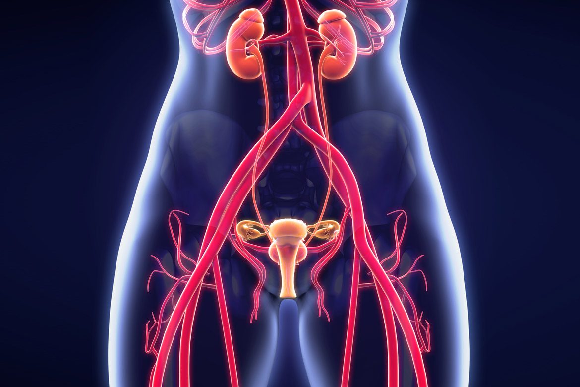
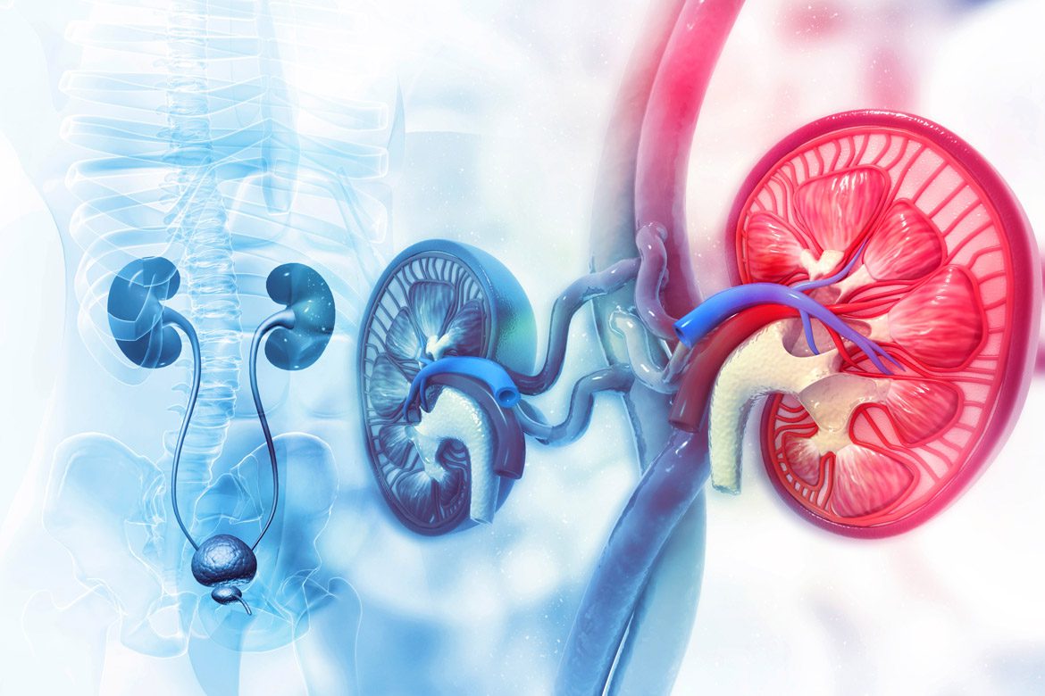
In addition to his focus on prostate health, Dr. Shahait provides comprehensive care in Orange County, CA for a wide range of general urology conditions, including flank pain, kidney stones, hematuria (blood in the urine), and urinary tract disorders. His expertise as a kidney disease specialist spans both medical and surgical management, offering personalized treatment plans to improve patient outcomes. With a commitment to cutting-edge technology and patient-centered care, Dr. Shahait continually integrates the latest advancements in urology to deliver high-quality, evidence-based treatments tailored to each patient’s needs.
Conditions We Treat


Testimonials
I highly recommend Dr. Shahait, what an amazing person....From the initial phone call to the Surgery and Post surgery he has been very professional and a genuine person who cares about his patients. Thank you so much and I feel 100% better now.
-Y.P., courtesy of HealthFinder
Dr. Shahait is a very caring and professional doctor. He works with a full conscious. Dr. Shahait gives the patient all the time needed and gives a detailed explanation of the case and the way it should be addressed, cared for, and treated. I strongly recommend Dr. Shahait!
-N.E., courtesy of HealthFinder
We received great care from Dr. Shahait. He was patient, thorough, cautious and competent. He was very responsive and kind and I would highly recommend him.
-R.A., courtesy of HealthFinder
Words can never describe how humble, honest, caring, and precise Dr. Mohammad is. He has the greatest role in my Dad's fighting cancer journey. Will never forget how he stood by our side and been more than just a doctor.
-F.S., courtesy of HealthFinder